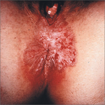
The biopsy revealed that the patient had Paget’s disease of the vulva. Paget’s disease of the external genitalia is an uncommon primary cutaneous adenocarcinoma of apocrine gland-bearing skin. Lesions present as geographic red macules that often appear excoriated or have an eczematoid appearance. Lesions may be dotted with small, white patches. Patients may also present with erythematous, eczematous, or leukoplakic plaques.
The most commonly involved site is the vulva, although perineal, perianal, scrotal, and penile skin may also be affected. Aside from the location, Paget’s disease of the vulva is morphologically and histologically identical to Paget’s disease of the nipple.
Up to 25% of patients with genital Paget’s disease have an underlying neoplasm. Associated malignancies include carcinomas of Bartholin’s glands, urethra, bladder, vagina, cervix, endometrium, and adnexal apocrine tissue. Only a small number of cases represent a direct extension of an underlying carcinoma.
In this case, the patient was sent to a gynecologic oncologist for a wide local excision of the involved area. The health care team planned to follow the patient closely to detect any recurrence at an early stage.
Photos and text for Photo Rounds Friday courtesy of Richard P. Usatine, MD. This case was adapted from: Mayeaux EJ. Paget disease of the external genitalia. In: Usatine R, Smith M, Mayeaux EJ, et al, eds. Color Atlas of Family Medicine. 2nd ed. New York, NY: McGraw-Hill; 2013:514-518.
To learn more about the Color Atlas of Family Medicine, see: http://www.amazon.com/Color-Family-Medicine-Richard-Usatine/dp/0071769641/
You can now get the second edition of the Color Atlas of Family Medicine as an app by clicking on this link: http://usatinemedia.com/