Compare the affected and uninjured extremities
Inspect, palpate, and assess the active and passive range of motion, strength, and neurovascular status of both arms, with the uninjured side serving as a comparison. The scapula, shoulder, and wrist are also involved in throwing, so these joints should be examined along with the elbow.
Measure range of motion. Normal ranges for the flexion-extension arc are 0 to 140°, with 75° of pronation and 82° of supination.12 Use a goniometer, if available, to ensure accuracy and reproducibility,1 and pay close attention to the position that elicits pain.
In medial epicondylitis, the full range of motion should be preserved. Patients experience pain at the medial epicondyle and overlying flexor-pronator mass proximately, and pain or weakness with resisted wrist flexion, and resisted pronation, at full extension.4,11 Flexor-pronator strain will produce similar findings, but edema or ecchymosis may be present and there may be pain immediately distal to the medial epicondyle.11
Pain associated with injury to the UCL—which courses distal and slightly posterior to the medial epicondyle—typically occurs 2 cm distal to the medial epicondyle over the anterior bundle. Tenderness over the UCL has a sensitivity of 81% to 94% for UCL tears, but a specificity of only 22%.13
Physical maneuvers can help identify source of elbow pain
A complete UCL tear can cause valgus gapping as small as 3 mm, which makes it difficult to detect on physical exam alone.4 Orthopedic and sports medicine literature recommend that 3 maneuvers be used to identify UCL pathology:4,14,15
The valgus stress test (FIGURE 1) assesses the effects of valgus stress on the UCL. Gapping >3 mm signifies UCL instability. The test has a sensitivity of 66% and a specificity of 60% for detecting a UCL strain or tear.13,16
The milking maneuver (FIGURE 2), performed by the patient (or by a clinician if the patient lacks flexibility), reproduces a common pitching exercise. Medial elbow pain or apprehension indicates UCL injury.13,16
The moving valgus stress test (FIGURE 3A-C) is done in an effort to recreate the flexion angles of the elbow during the late cocking and early acceleration phases of throwing. Pain anywhere in the arc of motion suggests a UCL injury; pain elicited at 45° of flexion suggests osteochondrosis of the humeral capitellum, while pain closer to full extension suggests osteochondrosis of the trochlea.13,16
FIGURE 1
Valgus stress test
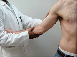
With the injured elbow at 30° of flexion, the shoulder abducted and fully externally rotated, and the patient’s wrist under your arm, place one hand laterally over the elbow. Place the other hand under the ulna and the thumb over the ulnar collateral ligament and apply valgus stress. Gapping >3 mm is abnormal.
FIGURE 2
Milking maneuver
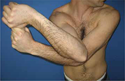
The patient grasps the thumb of the affected arm and pulls downward, with the affected elbow positioned as shown, stressing the ulnar collateral ligament (UCL). Elbow pain or apprehension is positive for UCL injury.
FIGURE 3
Moving valgus stress test
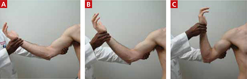
With the shoulder in abduction and maximum external rotation (A), place the elbow in maximum flexion and apply valgus force (B), and extend the elbow from full flexion to full extension (C) in an attempt to reproduce the medial pain.
Does your patient have 2 positive valgus tests and posterior pain?
Valgus extension overload syndrome, which is caused by repetitive stress and results in osteophytes, chondromalacia of the medial olecranon fossa, tension in the UCL, and compression of the radiocapitellar joint, will also produce positive valgus stress and positive moving valgus stress tests. Keep in mind, however, that patients with valgus extension overload often have loss of full extension and posterior elbow pain with forced elbow hyperextension.17
Look for ulnar nerve injury
The physical examination should also be used to test for ulnar nerve injury. The elbow flexion test—a provocative maneuver in which the patient flexes the elbow as far as possible and reports any tingling or numbness of the hand—should be included in the work-up. Symptoms that develop in <60 seconds indicate a positive test for ulnar nerve compression, with the pinky and ulnar half of the ring finger most likely to have loss of vibration and light touch perception. A positive Tinel’s sign over the cubital tunnel is an indication of ulnar neuritis.18