Diagnosis: Adenocarcinoma
The chest x-ray revealed a massive right-sided pleural effusion resulting in a hemothorax, tense mediastinum, and partial collapse of the left lung. A CT scan (FIGURE 2) of the chest revealed a 5-cm mass in the right lung. Thoracocentesis was performed and 11 liters of pleural fluid were removed.
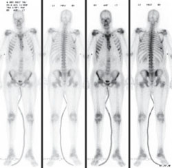
Cytological examination of the pleural fluid revealed adenocarcinoma of the lung. A diagnosis of adenocarcinoma of pulmonary origin was supported by immunohistochemical tests that were positive for thyroid transcription factor 1, cytokeratin 7, carcinoembryonic antigen, and epidermal growth factor receptor (EGFR), which made findings from other sites of adenocarcinoma less likely. A bone scan revealed metastases to the sixth rib and sternum, indicating stage IV adenocarcinoma (FIGURE 3).
FIGURE 2
Mass in lung
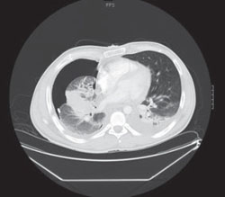
FIGURE 3
Bone scan reveals metastases
A cancer that presents at advanced stages
Lung cancer is the leading cause of cancer death in the United States in both men and women. The National Cancer Institute estimates that 159,390 people will die of lung cancer this year.1
More than 219,000 cases of lung cancer will be diagnosed this year;1 primary adenocarcinoma, a subtype of non-small cell lung cancer (NSCLC), is the most commonly diagnosed form of lung cancer.2 Most NSCLC cases will present at advanced stages, which limits treatment options and leads to a poor prognosis.
Cigarette smoking remains the most significant risk factor for lung cancer and, according to the American Cancer Society, smoking is responsible for at least 30% of all cancer deaths.3 Moreover, the US Department of Health and Human Services has found that 80% of lung cancers are attributable to smoking.4