Diagnosis
Immunohistochemistry was positive for CD1a, Langerin, and CD163.
LCH is a rare hematopoietic disorder that consists of the proliferation of monocyte-derived dendritic cells or macrophages. It is one of several histiocytic disorders, which are typically subdivided into LCH versus non-LCH disorders. This delineation arises from the molecular components found in LCH, including Langerin-positive, CD1a-positive, and S100-positive dendritic cells. The presentation of LCH can vary from solitary cutaneous lesions to disseminated, systemic disease. Systemic LCH can affect nearly any organ. High risk lesions are located in the liver, spleen, or bone marrow. Low-risk lesions are located in the bone, lung, lymph nodes, gastrointestinal tract, and CNS.
 University of Miami
Dr. Angelina Labib
The prognosis of LCH is usually dependent on the extent of involvement; LCH is usually risk stratified by location and extent of disease. The pathogenesis of LCH is not entirely known; however, it is thought to be secondary to a neoplastic, proliferative process arising from activation of the mitogen-activated protein kinase pathway. Certain mutations have been identified as the activators of this pathway, including BRAF-V600E, which is thought to be a fusion mutation that up-regulates the activity of MAPK.
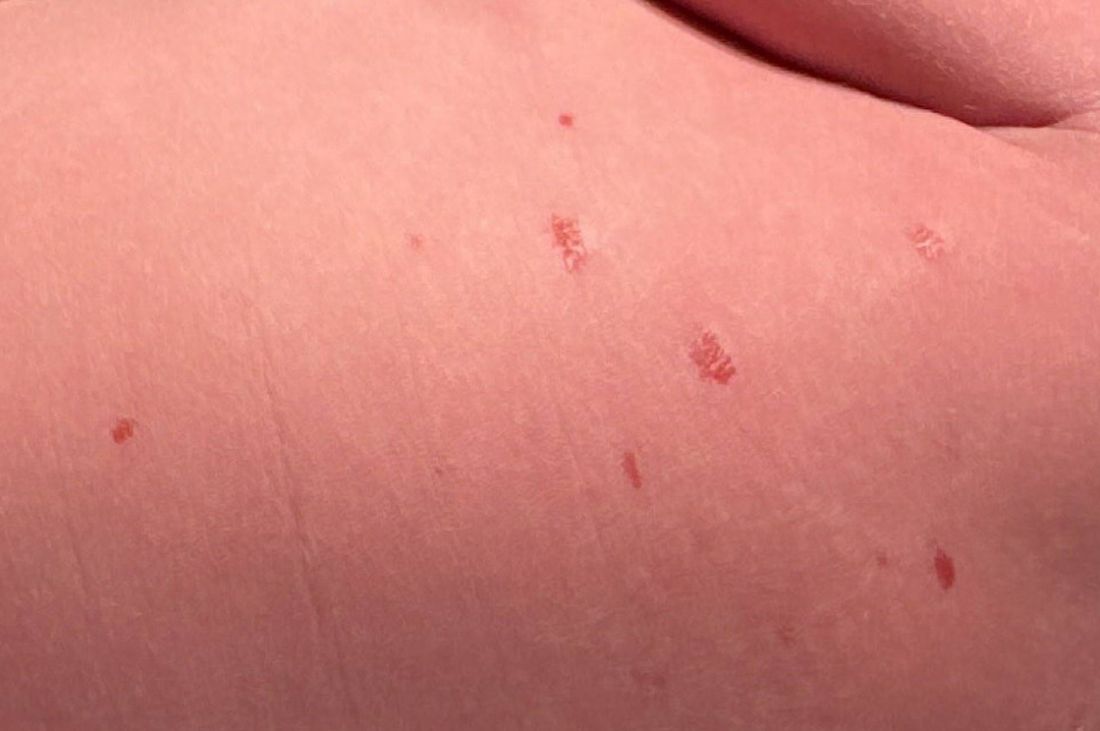
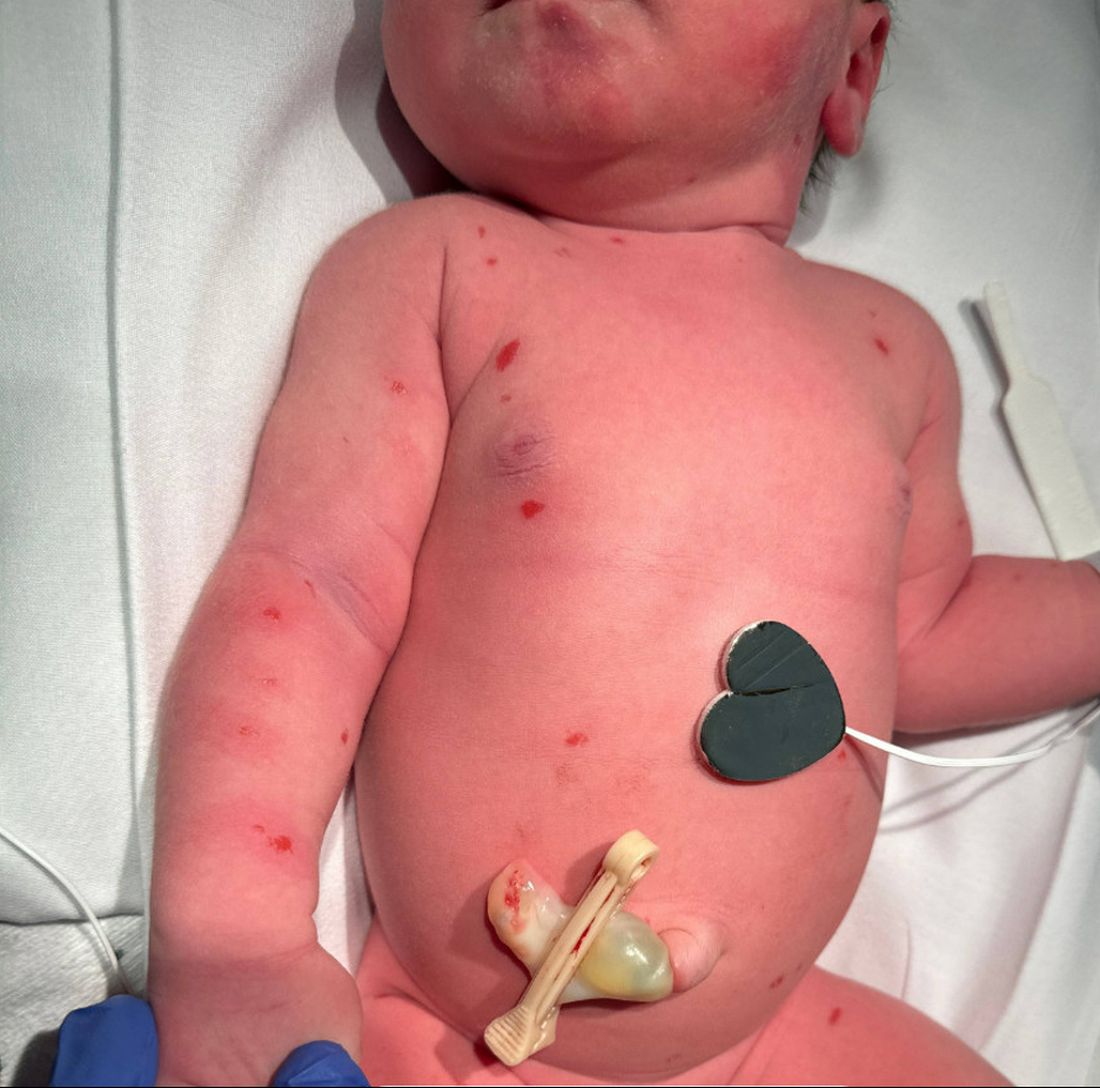
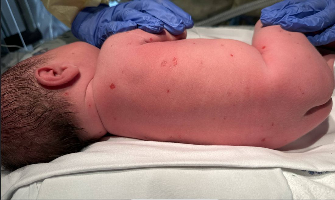
Congenital self-healing reticulohistiocytosis is a term that has been used for a benign variant of LCH that appears to be self-limiting. It typically appears at birth or during the neonatal period and presents as an isolated lesion or more commonly multiple hemorrhagic, vesicular, and crusted papules. Typically, the lesions begin to regress spontaneously within weeks to months. Work-up should include imaging and laboratory monitoring to preclude systemic involvement of the abdomen, skeletal system, and bone marrow. Often no treatment is required for what has been termed the “self-healing” type. Patients must be monitored for recurrence as it is clear that cases that appear to be self-healing may have recurrence with systemic progression or be associated with later findings of diabetes insipidus.
 MiLo Studios
Xochitl Longstaff
In this case, the infant initially presented with erythematous crusted papules, some hemorrhagic and others purpuric, throughout the trunk and extremities which began to resolve almost immediately with biopsy findings consistent with LCH. To ensure that there was no extracutaneous involvement, the patient underwent imaging of the abdomen via ultrasound and a skeletal survey via x-ray. After confirming that the disease was limited to cutaneous findings, the patient was discharged from the hospital with close follow-up with dermatology and hematology/oncology. Genetic testing of the biopsy returned positive for the BRAF-V600E mutation. There has been no evidence of recurrent skin lesions nor systemic signs or symptoms over the succeeding 8 months.
The maternal history of varicella zoster virus and shingles alerted clinicians to consider varicella zoster infection as a potential diagnosis. The presence of shingles in a pregnant woman, as compared with primary zoster, is associated with prior establishment of serologic status that is generally protective in utero, and in this case it was clarified that the shingles event was not during pregnancy. Primary varicella in utero can cause congenital varicella syndrome with exposure in the first trimester.
 University of California, San Diego
Dr. Lawrence Eichenfield
Herpes simplex virus (HSV) infection can present with localized or generalized vesicular erosive and/or crusted lesions. Evaluation of a newborn with such lesions should include polymerase chain reaction tests of cutaneous lesions and/or viral culture, more extensive work-up as clinically indicated. This child was placed on acyclovir while awaiting diagnostic study results. Diagnostic testing, specifically the pathologic findings on skin biopsy were diagnostic of LCH.
Scabies is unlikely in this setting given the presence of lesions at birth and the lack of exposure to a possible parasite. Scabies infection is typically characterized with burrows and crusted nodules that appear around web spaces of the hands and feet and is not seen congenitally.
The rash does not resemble a “blueberry muffin rash,” which is characteristic of congenital rubella, other congenital infections, and rarely other conditions and is associated with extramedullary hematopoiesis. These lesions may appear as purpuric papules and nodules, and generally not vesicles.
Dr. Angelina Labib is the Post-Doctoral Pediatric Clinical Research Fellow at UCSD Rady Children’s Hospital in San Diego, California. Xochitl Longstaff is currently completing a research year as a Pediatric Clinical Research Fellow at UCSD Rady Children’s Hospital prior to finishing her final year at the David Geffen School of Medicine at the University of Californa, Los Angeles. Dr. Eichenfield is vice chair of the department of dermatology and professor of dermatology and pediatrics at the University of California, San Diego, and Rady Children’s Hospital. .
Suggested Reading
Allen CE et al. Blood. 2015 Jul 2;126(1):26-35. doi: 10.1182/blood-2014-12-569301.
Frade AP et al. AAn Bras Dermatol. 2017;92(5 Suppl 1):40-42. doi: 10.1590/abd1806-4841.20175308.
Kallesh A and Kumar VS. N Engl J Med. 2021 Dec 9;385(24):e86. doi: 10.1056/NEJMicm2112460.
Prose NS and Antaya RJ. Neoplastic and Infiltrative Diseases. In: Eichenfield LF ed. Neonatal and Infant Dermatology, 3rd Edition. Elsevier, 2015.