Jane Hwang, MD Emily Wong, MD San Antonio Uniformed Services Health Education Consortium, San Antonio, Tex (Dr. Hwang); Department of Dermatology, Scott Air Force Base, Ill (Dr. Wong) jane.hwang.1@us.af.mil
DEPARTMENT EDITOR Richard P. Usatine, MD University of Texas Health Science Center at San Antonio
The authors reported no potential conflict of interest relevant to this article.
Our patient’s history provided 2 important clues (one vascular, one not) to explain the painless ulcer that had been on his right shin for a year. A punch biopsy made the diagnosis clear.
A 63-year-old morbidly obese man presented to our clinic with a non-healing, slowly growing, painless ulcer on his right shin that he’d had for one year. It was not actively bleeding or draining, but the scab had come off one month earlier and the wound did not close. The patient denied any trauma to the area or foreign travel. Bacitracin and triamcinolone creams hadn’t helped.
Our patient’s medical history included diabetes, hypertension, hyperlipidemia, and worsening venous insufficiency. He was not currently using compression stockings, but they had helped him in the past.
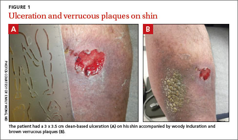
On examination, we noted a 3 x 3.5 cm well-demarcated, somewhat geometric, clean-based ulceration on the patient’s right medial shin (FIGURE 1A). There was no significant erythema, purulence, tenderness, warmth, or drainage of the ulcer. The base had seemingly normal granulation tissue. Woody induration, verrucous plaques, and confluent erythematous, violaceous, indurated patches were adjacent to the ulcer (FIGURE 1B). The patient also had severe pitting edema on his lower legs.
WHAT IS YOUR DIAGNOSIS? HOW WOULD YOU TREAT THIS PATIENT?