Grandhi R, Shamloul N, Powell M. Purpuric bullae on the lower extremities. Cutis. 2020;105:282, 286-287.
In the article above from the June 2020 issue, the images were incorrect. The correct images appear below. The article has been corrected online at www.mdedge.com/dermatology. We apologize for the error.
Quiz Images
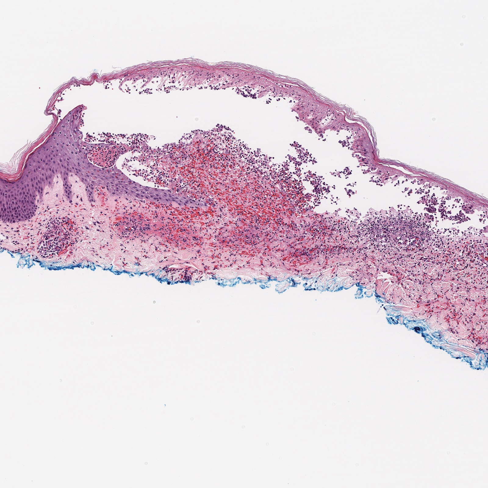
H&E, original magnification ×100.
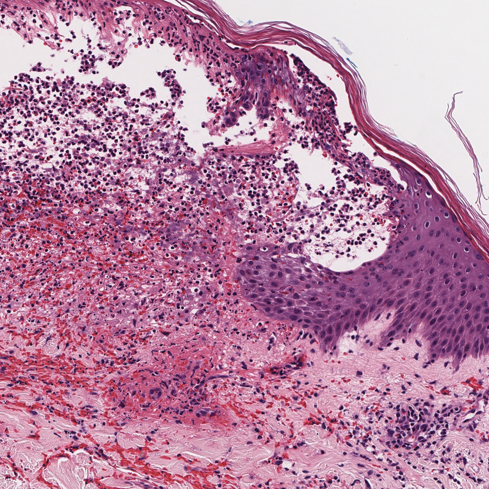
H&E, original magnification ×200.
Discussion Images
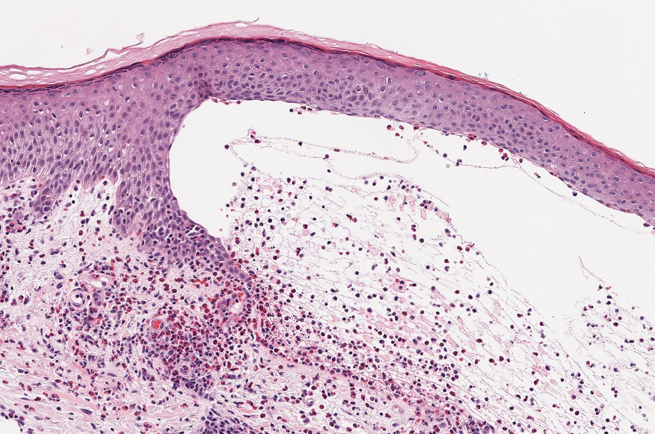
Figure 1. Bullous pemphigoid. Subepidermal bulla with eosinophils and neutrophils within the bulla as well as numerous dermal eosinophils (H&E, original magnification ×200).
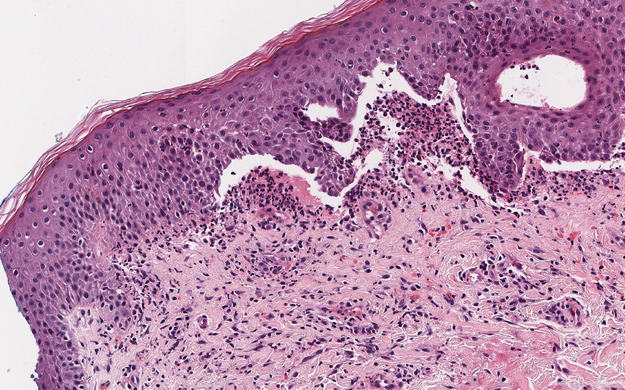
Figure 2. Linear IgA bullous dermatosis. Subepidermal bulla with numerous neutrophils within the bulla and sparse dermal eosinophils and neutrophils (H&E, original magnification ×200).

Figure 3. Stasis dermatitis. Pauci-inflammatory subepidermal bulla with fibrin. The overlying epidermis is intact. The dermis shows cannon ball angiomatosis, red blood cell extravasation, and fibrosis (H&E, original magnification ×200).
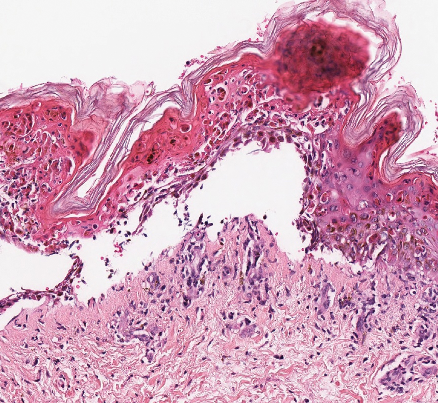
Figure 4. Stevens-Johnson syndrome/toxic epidermal necrolysis. Pauci-inflammatory subepidermal separation with acute epidermal necrosis. There is minimal dermal inflammation and pigment incontinence (H&E, original magnification ×200).