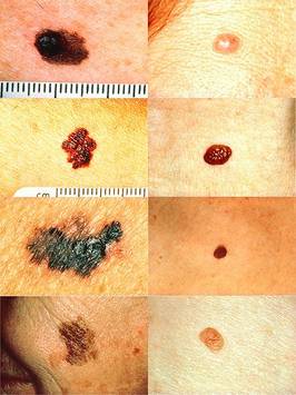
 Images from National Cancer Institute via Skin Cancer Foundation, merged by WikiMedia Commons user Stevenfruitsmaak
Left side, top to bottom: melanomas showing (A) asymmetry, (B) irregular border, (C) unusual coloring, and (D) diameter that had changed in size. Right column: normal moles.
The month of May marked 25 years since dermatologists began using the ABCD rule to help diagnose melanoma, and advances in diagnosis since then have leaned toward newer and better use of imaging technology instead of clinical mnemonics.
Advances over the past quarter-century have focused on helping physicians “see” melanoma better. Dr. Darrel Rigel, of New York University Medical Center, described some of these at the annual meeting of the American Society for Mohs Surgery.
Dermoscopy allowed non-invasive imaging of melanomas. Digital photography came along, and some dermatologists began using serial digital imaging to track changes in moles. Most recently, various groups have been trying to add assessments using infrared (non-visible) light through computer-aided programs to aide diagnosis.
As a woman of letters, I have to admit a fondness for the ABCDs. But the imaging advances do have more of a new-school, digital-age feel to them. I think both will happily coexist in dermatology. Time will tell if that’s old-school thinking as the digital age advances.
– Sherry Boschert